Uncovering the human brain by recognizing distinct regions with the purpose to derive personality aspects (e.g., criminality), electrochemical impluses percolating through a large number of neurons, or the visualization of nerve bundles based on Magnetic Resonance Imaging (MRI) data stays as top research area in the last centuries. The following gallery shows a historical timeline beginning from 1583 up to 2017 including advancements in the discovery of human brain structures.
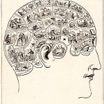
The German physician Georg Bartisch published a book on ophtalmology containing a woodcut of several overlays to reveal the layers of the brain’s anatomy in 1583. The content of his book addresses the anatomy of the head and eye, then moves on to discuss treatments for cataracts, trauma, strabismus, and afflictions presumed to be caused by witchcraft. Toward the eighteenth century, researchers studied the skull itself with the hypothesis that the shrunken areas of the skull correspondence to the area of the brain as contrary perspective to the inner explanation of organic structures. The promise was to detect areas that refer to a particular personality trait, character, or behavior. The field of phrenology was born during this time period and the “Phrenological Chart of the Faculties” (1883) shows twenty-seven regions (e.g., the sense of form and size) based on the shape of the skull. The number of regions increased by including all types of intellectual, mental, and emotional aspects. The field of phrenology was only a beginning to describe the modularity of the mind but the human brain is even more complex when it comes to analyze their biological structure.
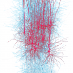
Modern neuroscientists initiated the Blue Brain project to analyze the adult human brain with over one hundred billion neurons in 2008. The projects aims to build digital reconstructions (computer models) of the brain with an unprecedented level of biological detail. Supercomputer-based reconstructions and simulations are used to exploit interdependencies in the experimental data to obtain dense maps of the brain. A computer-generated model produced with IBM’s Blue Gene supercomputer shows the thirty million connections between ten thousand neurons in a single neocortical column. This example shows the disruptive shift from identifying regions to information flows between neuron connections. But this significant breaktrough is representing only a small slice of the brain cortex. Visualizing the entire brain requires enormous scalability capacities.
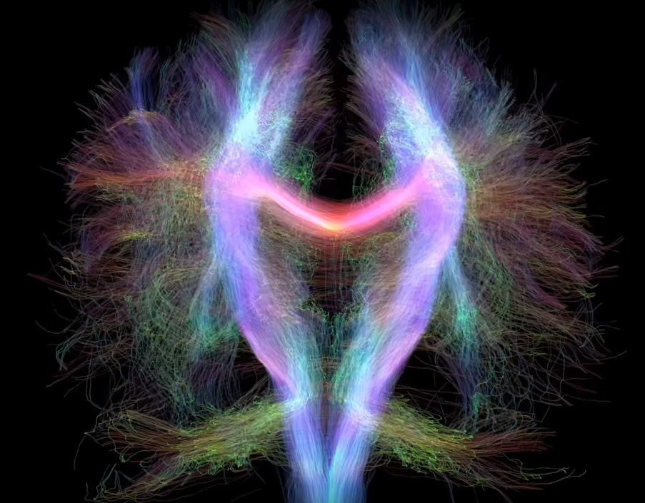
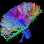
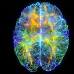
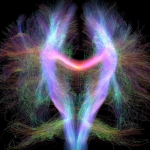
For this purpose, the Human Connectome Project was established to construct a map of the complete structural and functional neural connections in 2010. This requires advanced high-field imaging technologies and neurocognitive tests to map the human connectome. As a result, 3D models can be explored interactively. This allows the detailed analysis of diseases like Alzheimer’s disease or dementia as well as anxiety or depressive disorders. Recognizing different structures of individuals was enabled through the combination of neuroimaging technologies that are able to generate MRI/EEG data with rendering technologies like the Unity 3D game-engine powered by NVIDIA’s GPU computing. The Glass Brain (2015) allowed the exploration of brain networks and their activity in real-time. Those MRI data are generated by a magnetic field surrounding the body part to be scanned, and sends radio waves into the body. The increasing amount of data and their visual analysis require huge hardware capacities. The Laboratory of Neuro Imaging (LONI) houses a large super computer, over 50 workstations and a data archival system of over 100 terabytes, with the goal to analyze MRI data. This is even necessary to analyze fiber nerve bundles that was published with a MRI data visualization on the 27th of January 2017.
Those advancements have extended the community to a diverse and multidisciplinary research area that gathers experts around neuroscience, psychology, medical science, and computer science. The historical examples have shown the major direction to a deeper discovery of the brain regarding their anatomical structure and dynamic activity. But also their required capacities (e.g., neuroimaging technologies, supercomputer infrastructures, and visual analysis tools) must scale up with the exploration process by uncovering patterns of the human brain.
